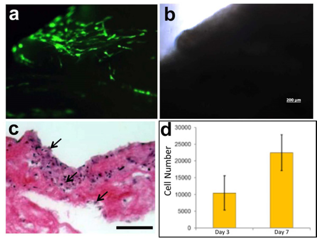
Figure 4. Cell growth on acellular porcine pancreas.
(a) Fluorescent and bright field (b) images of GFP-labeled human amniotic fluid-derived stem cells (hAFSC) cultured on porcine acellular pancreatic matrix shown 48 hours after seeding. (c) H&E staining of sections of acellular porcine pancreas seeded with hAFSC 7 days after seeding (scale bar = 100µm). (d) Proliferation of hAFSC seeded onto acellular porcine pancreas, measured by MTS assay, shows increase in cell number from day 3 to day 7. Values expressed as mean ± stdev, n=4.

