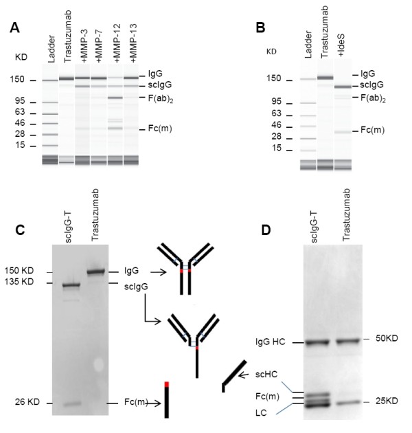
Figure 1.

Detection of trastuzumab proteolytic cleavage in vitro. (A) Capillary electropherograms (CEs) of trastuzumab and matrix metalloproteinase (MMP)-digested trastuzumab under nonreducing and denatured running conditions. (B) CE of trastuzumab and IgG-degrading enzyme S (IdeS)-digested single hinge cleaved trastuzumab (scIgG-T). (C) Purified scIgG-T under nonreducing and denaturing running conditions and stained with Coomassie blue. (D) Trastuzumab and scIgG-T under reducing and denaturing running conditions and stained with Coomassie blue. Fab, fragment antigen binding; Fc(m), Fc monomer; HC, heavy chain; LC, light chain.
