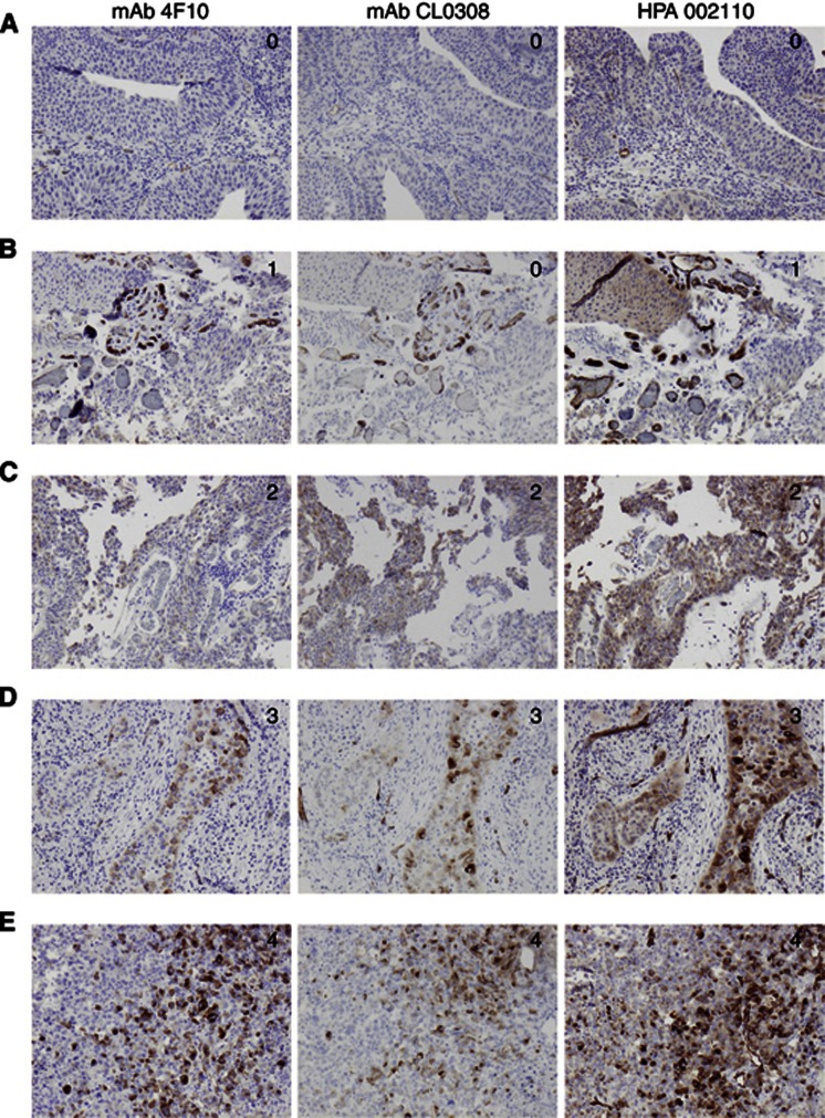
Figure 1.
Sample images of PODXL staining in urothelial bladder cancer—comparison of three different antibodies. Immunohistochemical images ( × 10 magnification) of five cases (A–E) with tumours denoted as having (0) negative, (1) weak, (2) moderate, (3) partly (<50%) membranous and (4) membranous PODXL expression in >50% of tumour cells, using three different antibodies.

