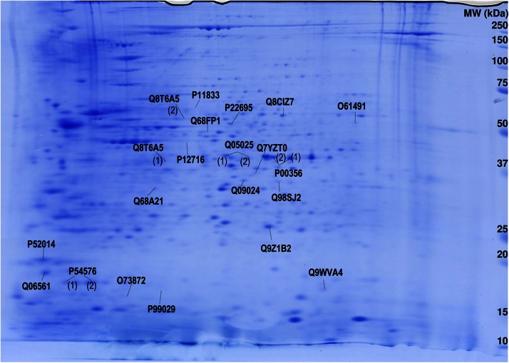
Figure 1.
Two-dimensional gel map of Aplysia abdominal ganglia proteins with statistically significant different levels after serotonin treatment. Aplysia abdominal ganglia proteins were extracted, and 700 μg were applied on an immobilized pH 3–10 non-linear gradient strip, followed by 9%–16% linear gradient polyacrylamide gel. Gels were stained with Coomassie Blue, spots were analyzed, and proteins were assigned using MASCOT software (14). UniProtKB accession numbers for protein identification are given.

