
Figure 2.
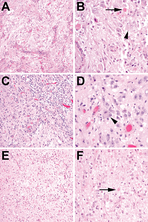
IDH1 wild‐type grade I gangliogliomas. (A,B) A 17‐year‐old male with a tumor in the right parietal lobe underwent resection 13 years ago, which revealed a ganglioglioma with binucleate neurons (arrowhead) and eosinophilic granular bodies (arrow). No further recurrences were reported. (C,D) A right temporal lobe mass from a 10‐year‐old male showed ganglion cells with binucleation (arrowhead) admixed with bland‐appearing glial elements. Even after gross total resection the tumor recurred 2 years later (E,F) but was still recognizable as a grade I ganglioglioma, with abundant ganglion cells and scattered eosinophilic granular bodies (arrow). Follow‐up since then (3 years after first recurrence) has shown no evidence of additional recurrences.