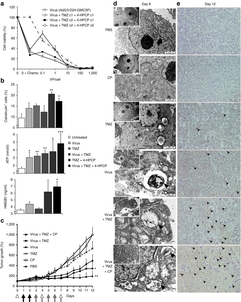
Figure 1.
Immunogenic cell killing and increased autophagy coincide with tumor growth inhibition in oncolytic adenovirus, temozolomide (TMZ)- and cyclophosphamide-treated prostate cancer. (a) Cell-killing efficacy of combination treatments. Ten virus particles (VP)/cell of oncolytic adenovirus combined with TMZ (c1 = 0.035, c2 = 0.105 mg/ml) and 4-HPCP (c1 = 0.003125, c2 = 0.00525 mg/ml) resulted in superior cell killing over chemotherapeutic agents or virus alone (P < 0.01, P < 0.05, respectively). (b) Immunogenicity of cell death. Combination treatment with 100 VP/cell of Ad5/3-D24-GMCSF virus, TMZ (0.0025 mg/ml), and 4-HPCP (0.00208 mg/ml) resulted in significant increase in calreticulin-positive PC3-MM2 cells, and extracellular ATP and HMGB1 levels, as compared with untreated cells. Treatment with TMZ and 4-HPCP increased calreticulin-positive cells and ATP release, whereas oncolytic virus seemed to induce mostly ATP and HMGB1 release, but lacked significant induction of others. Data in a,b are representative of three independent experiments. (c) Efficacy of combination therapy in vivo. Nude/NMRI mice bearing subcutaneous PC3-MM2 xenografts were treated intratumorally with Ad5/3-D24-GMCSF virus or growth medium (black arrows) followed by intraperitoneal injections of TMZ or saline (gray arrows), and cyclophosphamide (CP) or saline (white arrowheads). Virus + TMZ, and virus + TMZ + CP treatments significantly inhibited tumor growth as compared with phosphate-buffered saline (PBS) control. (d,e) Induction of autophagy after combination therapy in vivo. (d) Electron microscopy on fixed tumor tissues revealed large tumor cells with enlarged nuclei, abundant mitochondria, ribosomes, and glycogen deposits (gray vacuoles). Autophagosomes and autolysosomes were only found in virus-, virus + TMZ-, and virus + TMZ + CP-treated tumor cells (black arrowheads). VPs were observed inside some of the autophagic cells (white arrowheads). (e) Tumors were assessed by immunohistochemistry for LC3, a membrane-bound protein accumulating on late autophagosomes. Punctate staining pattern was considered indicative of autophagy (black arrowheads; ×40 original magnification). Error bars represent the mean ± SEM. All studies, n = 3–6; *P < 0.05; **P < 0.01; ***P < 0.001; unpaired t-tests in a,b; one-way analysis of variance repeated measures in c. 4-HPCP, 4-hydroperoxycyclophosphamide; ATP, adenosine triphosphate; GMCSF, granulocyte-macrophage colony-stimulating factor; HMGB1, high-mobility group box-1.

