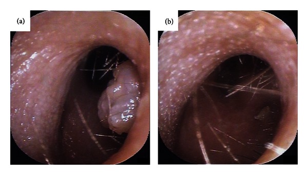
Figure 1.

Endoscopic photographs of the left external auditory canal. (a) Fibroepithelial polyp at the posterior wall of the inlet of the left external auditory canal. (b) Left external auditory canal 1 week after resection of the fibroepithelial polyp.
