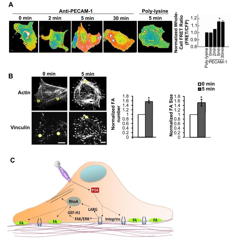
Figure 6. Local tensional forces on PECAM-1 elicit global RhoA activation and focal adhesion growth.
(A) ECs expressing the RhoA biosensor were incubated with poly-lysine or anti-PECAM-1-coated beads (4.5μm) and subjected to force with a permanent ceramic magnet for the indicated times. Cells were fixed and analyzed for FRET. Whole cell FRET ratios were calculated for each condition. Autofluorescent beads are highlighted in black dotted circles (n > 45 cells/condition from 4 independent experiments, *p < 0.05). (B) Adherent ECs on FN were incubated with anti-PECAM-1-coated magnetic beads and subjected to force for the indicated times. ECs were fixed stained with phalloidin and an anti-vinculin antibody to mark focal adhesions. Focal adhesion number and size were quantified using NIH ImageJ software. Values were normalized to the “No Force” condition. Location of the beads are highlighted in yellow circles (n > 30cells/condition from 3 independent experiments, *p < 0.05, scale bar = 10μm). (C) Model of PECAM-1-mediated mechanotransduction. Local tensional forces on PECAM-1 results in global mechanosignaling and changes in cytoskeletal architecture. (See also Fig. S4)

