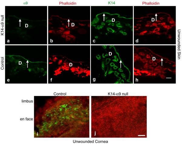
Figure 2. Effective knockout of α9 by K14-Cre mediated recombination in skin and cornea.
Unwounded skin (a–h) or cornea (i and j) were harvested either from K14-α9 null or control mice. Harvested skin was oriented with the epidermis on the top and panniculus carnosus below. Unwounded consecutive skin sections of K14-α9 null mice (a–d) or control mice (e–h) were treated either with rabbit anti-α9 (a, b, e and f) or K14 (c, d, g and h), as a marker for keratinocytes. No positive staining was detected for α9 in the epidermis (arrow) of K14-α9 null mice (a) compared to the positive staining observed for α9 in the control mice epidermis (e). Panels b, d, f and h depict phalloidin staining of their corresponding panels a, c, e and g respectively. Using whole mount confocal imaging, in the cornea, we confirm loss of integrin α9 staining at the limbus of cornea in K14-α9 null mice (i) compared to control mice (j). Dermis denoted as `D'. N=3, scale bar: 50 μm in (h) and 40μm in (i and j).

