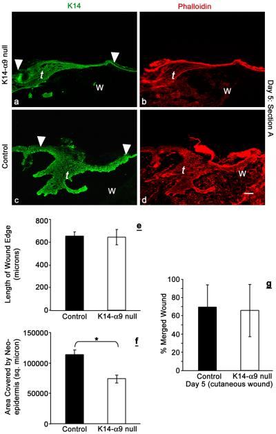
Figure 5. Poor re-epithelialization in K14-α9 null wounds compared to control mice.
Excisional cutaneous wounds were harvested at intervals of 5 d post-wounding. Harvested wounds were oriented with the epidermis on top and panniculus carnosus, below. Lengths and areas of the migrating tongues (t) were visualized by K14 (a and c) or phalloidin (b and d) staining. Graph represents average length of the wound edge (e) and area covered by the neo-epidermis (f). No statistically significant difference was observed in the length of the migrating tongue, whereas reduced area was covered by the tongue of K14-α9 null wound (*p < 0.001). The graph (g) shows the no difference in the distance traveled by the keratinocytes to cover the wound in microns. N=6, scale bar: 50μm.

