Fig. 7.
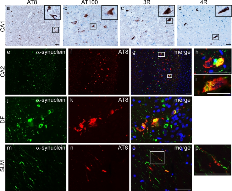
Tau expression and co-localisation with α-synuclein. Representative images from CA1 probed using immunohistochemistry with phospho-tau antibodies: AT8 (a) and AT100 (b). Inclusions contained a mixture of 3-repeat (c) and 4-repeat (d) tau isoforms. Double immunofluorescence images of CA2 (e–i) and DF (j–l) probed for α-synuclein (green) and AT8 (red) show strong co-localisation in a subset of neurons. AT8 immunoreactivity is detected on the dendritic processes of DF granule cells which express α-synuclein in the stratum lacunosum-moleculare (SLM, m–p). DAPI nuclear stain (blue). Scale bars represent 50 μm (b–d are at the same magnification as are e–g, j–o)

