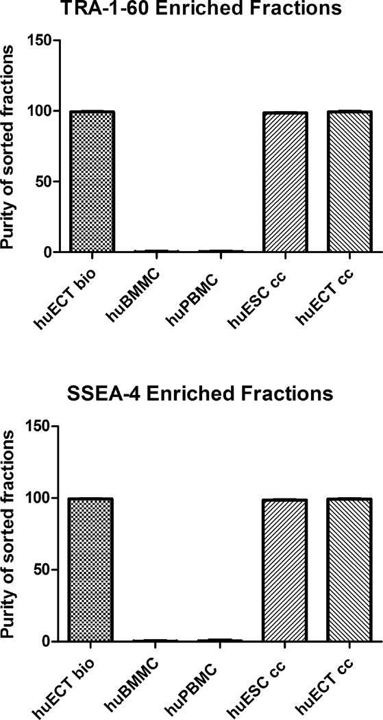
Figure 3.
The cells were biopsied directly from the primary tumors of the patients diagnosed with the embryonal carcinoma of the testes (huECT bio) (in the fig. 3, the patients encoded 001–009), the mononuclear cells from bone marrow (huBMMC) and from peripheral blood (huPBMC), the cultured human embryonic stem cell lines (H1, H13, H14) (huESC cc), and the cultured cells from metastasis to lungs of the testicular embryonal carcinoma (NT2D1) (huECT cc), labeled with the superparamagnetic Fvs targeting TRA-1–6- and SSEA-4, and isolated with magnetic sorter to enrich the samples’ purity better than 99.5% with the statistical significance accepted at p < 0.001.

