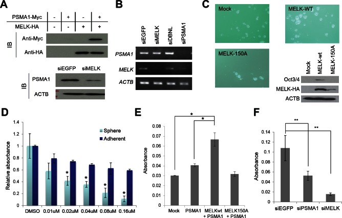
Figure 6. PSMA1 enhanced the mammosphere formation through the phosphorylation by MELK.
(A, B) PSMA1 protein was stabilized through the phosphorylation by MELK in breast cancer cells (A) although transcriptional level of PSMA1 was unchanged in the cells in which MELK expression was knocked down (B). (C) Wild-type MELK (MELK-wt) or kinase-dead mutant MELK (MELK-150A) expression vector was transfected into MCF-7 cells which were seeded onto an ultra-low attachment culture plate. The formation of mammosphere was enhanced in cells in which MELK-wt was transiently introduced than those transfected with mock vector or MELK-150A. The expression levels of one of cancer stem cell markers, Oct3/4, are shown. The cells which transiently over-expressed MELK-wt induced Oct3/4 expression while those transfected with mock vector or MELK-150A revealed no Oct3/4 expression. (D) OTSSP167 suppressed more significantly the formation of mammosphere than the growth of attached MCF-7 cells. The cells were plated onto ultra-low attachment culture plate or normal culture plate without or with OTSSP167 of given concentrations (*p<0.05, student's t-test). (E) The MCF-7 cells in which both PSMA1 and wild-type MELK (MELK wt) were co-overexpressed revealed higher number of mammosphere formation than the parental MCF-7 cells or those transfected with PSMA1 alone or PSMA1 + kinase-dead MELK (MELK D150A) (*p<0.0001, student's t-test). (F) The mammosphere formation of MDA-MB-231 cells, in which PSMA1 was knocked down, was suppressed (**p<0.05, student's t-test). Absorbance measured at 490 nm is indicated using that at 630 nm as a reference with a microplate reader. Error bars represent means ± SD of triplicates for experiments D-F.

