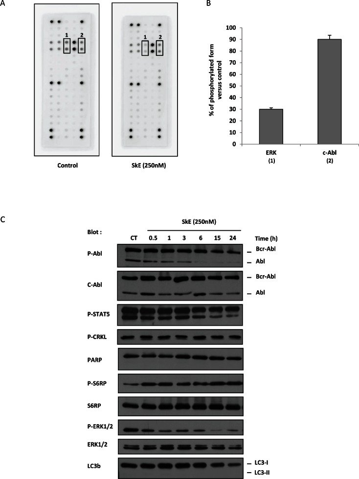
Figure 2. SkE treatment impairs ERK1/2 phosphorylation.
(A) K562 cells were treated with 250 nM of SkE for 2 h; then, cells were lysed, and cell lysates were loaded on a Pathscan multikinase® membrane. (B) Histograms represent the relative intensity quantification of the most regulated dot with Image J software. Results are expressed as the percentage of kinase phosphorylation in SkE-treated cells versus control cells. (C) K562 cells were incubated at 37°C with 250 nM SkE for the indicated times. Whole-cell lysates were prepared, and the expression of Phospho-C-Abl, C-Abl, Phospho-STAT5, Phospho-CRKL, PARP, Phospho-S6RP, S6RP, Phospho-ERK1/2, ERK1/2 and LC3b was visualized on a Western blot.

