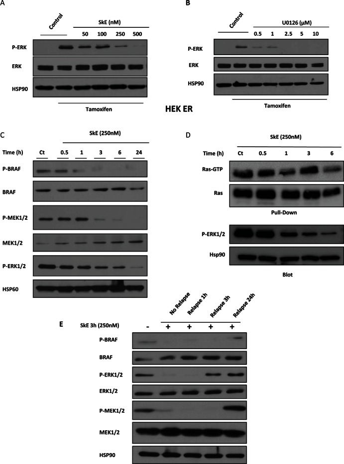
Figure 3. SkE can inhibit the RAF/MEK/ERK signaling pathway.
HEK Raf-ER cells were pre-treated with increasing doses of SkE (A) or U0126 (B) for 1 h. Cells were then treated with tamoxifen (1 μM) for one additional hour. Protein samples were separated by electrophoresis, and the expression of Phospho-ERK and ERK was visualized on a Western blot. (C) K562 leukemic cells were treated for different times with 250 nM SkE. The status of phosphorylation of BRAF, MEK and ERK was visualized by Western blot. (D) K562 cells were incubated at 37°C with 250 nM SkE for the indicated times. Ras activity was determined after GST-pull-down. Ras-GTP levels were determined using GST-c-Raf RBD to pull down active GTP-bound Ras from cell extracts by glutathione beads. The beads were washed 4 times and subjected to SDS/PAGE (12% polyacrylamide). Ras and Phospho-ERK1/2 proteins were detected by Western blot analysis. (E) K562 cells were treated with 250 nM SkE for 3 h. Cells were then washed and placed in fresh medium for 1 h, 3 h or 24 h. The BRAF, MEK and ERK1/2 protein levels and their phosphorylation status were analyzed by Western blot. HSP60 and HSP90 were used as the loading controls.

