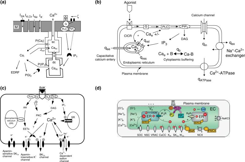
FIGURE 1.
Models of calcium dynamics in ECs. (a) A model introduced by Wong and Klassen.47 The model contained an internal calcium store that was divided into a superficial (sc) and deep (dc) compartments and four transmembrane currents: a sodium current (INa), a potassium current (IK), a Ca2+-activated potassium current (IK,Ca), and a stretch-activated calcium current (ISA). (b) A calcium dynamics model in human umbilical vein endothelial cells (HUVECS) introduced by Wiesner and coworkers.48 The model includes descriptions for CICR, CCE, kinetics for IP3 formations following thrombin receptor activation, and Ca2+ buffering. Formulations for the NCX and the PMCA pump were also included. (c) A model of endothelial cell electrophysiology presented by Schuster and coworkers (reprinted with permission from Ref 50. Copyright 2003 Springer). Model focuses on K+ currents from bradykinin-sensitive channels (i.e., the large-conductance BKCa channel, the apamin-insensitive small-conductance SKCa channel, and an NSC channel) and the apamin-sensitive SKCa channel that is not gated by bradykinin. (d) An endothelial cell electrophysiology/ionic dynamics model introduced by Silva et al.51 Model contains several transmembrane currents from: store-operated Ca2+ channels (SOC); nonselective cation (NSC) channels; voltage-regulated anion channel (VRAC); Ca2+-activated Cl– channels (CACC); inward rectifier K+ channels; Ca2+-activated K+ channels (small-conductance SKCa, intermediate-conductance IKCa); Na+-K+-ATPase (NaK) pumps; plasma membrane Ca2+-ATPase (PMCA) pumps; Na+/Ca2+ exchanger (NCX). The model accounts for changes in membrane potential (Vm) and in the intracellular concentrations of the four main ionic species following agonist induced IP3 release.

