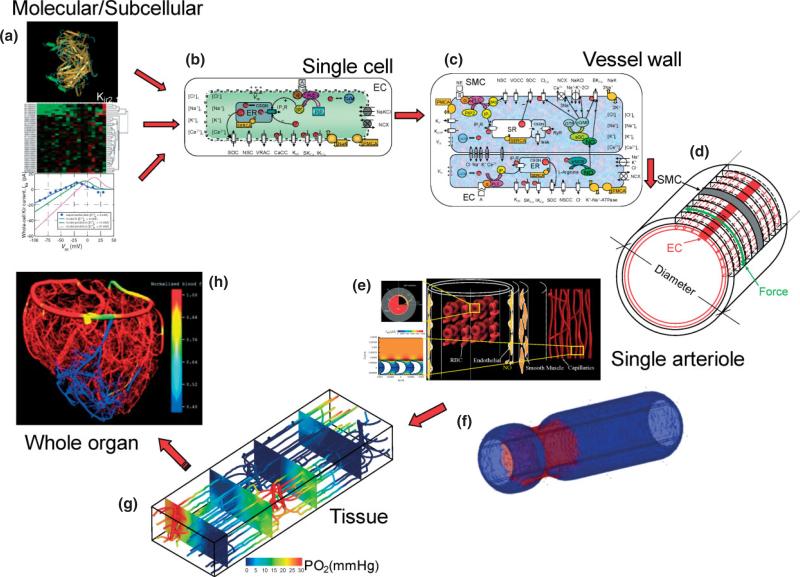
FIGURE 4.
Strategy for integrative modeling of the vasculature. (a) Genomic, proteomic and in vitro data such as electrophysiological recordings can be utilized to provide mathematical formulations for subcellular components and signaling pathways. Representative example shows the three-dimensional folding of KIR2.1 protein and the current–voltage behavior of a KIR channel in an EC. (b) An EC model51 that integrates such formulations and incorporates membrane channels, pumps and exchangers, intracellular compartments, and signaling mechanisms. The model can simulate membrane electrophysiology, dynamic behavior of Ca2+ and other ions, and the generation of second messengers and signaling factors. (c) EC and SMCs can be coupled through gap junctions and the diffusion of species like NO and IP3.78 (d) Multicellular models of the vascular wall can be constructed by placing EC and SMCs cells in an appropriate arrangement.79 (e) Example of a biotransport model investigating the diffusion of species (i.e., NO, O2) in and around a single arteriole and incorporates RBCs and nearby capillaries.80 (f) A biomechanics model of a single arteriole presents the constriction of the vessel at the site of norepinephrine application in the absence of longitudinal signal conduction. (g) Detailed computational model investigating blood flow and O2 distribution in three-dimensional vascular networks and mesoscale tissue volumes.95 (h) Reconstructed whole-organ vessel network and blood flow calculations.(Reprinted with permission from Ref 96. Copyright 2002 Society for Industrial and Applied Mathematics).

