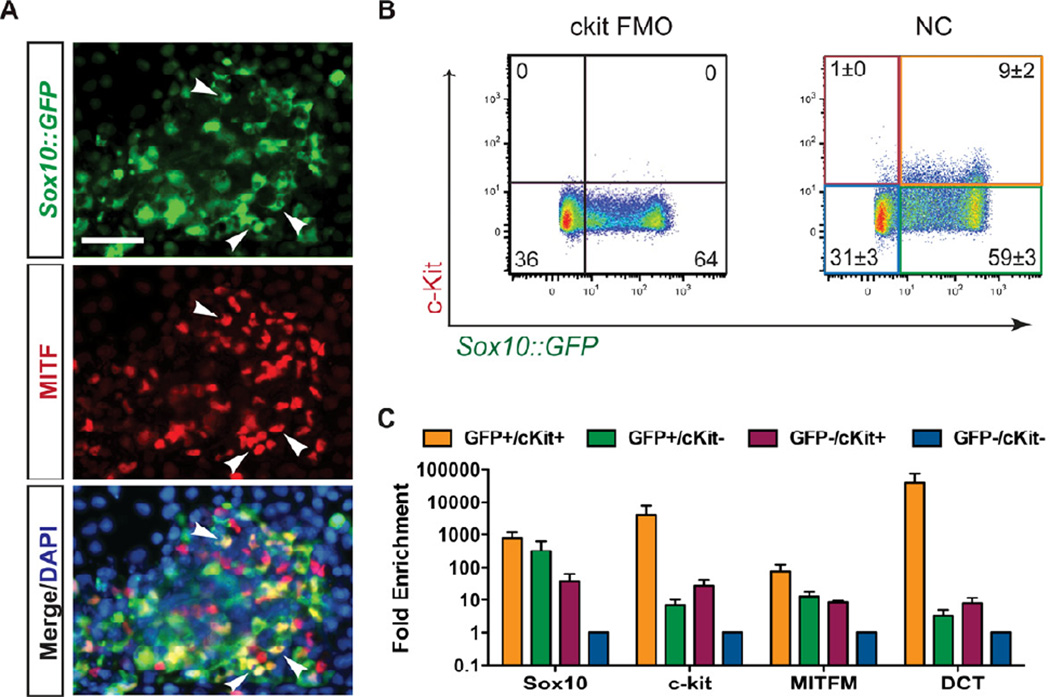
Figure 3. C-kit and Sox10 expression can be used to identify and isolate melanoblasts.
(A) Melanocyte progenitors can be identified in day 11 NC protocol-derived populations by coexpression of Sox10::GFP and the melanocyte transcription factor MITF (arrowheads). Scale bar represents 50µm. (B) Flow cytometry reveals the presence of a Sox10::GFP and c-kit co-expressing population. C-kit “fluorescence minus one” (FMO) was used a negative control for c-kit staining. (C) 4-way FACS sorting for Sox10::GFP and c-kit reveals an enrichment of the melanocyte markers MITFM and Dct in the SOX10/ckit double positive population by qRT-PCR. All error bars represent the s.e.m of at least three independent experiments. See also Figure S4.

