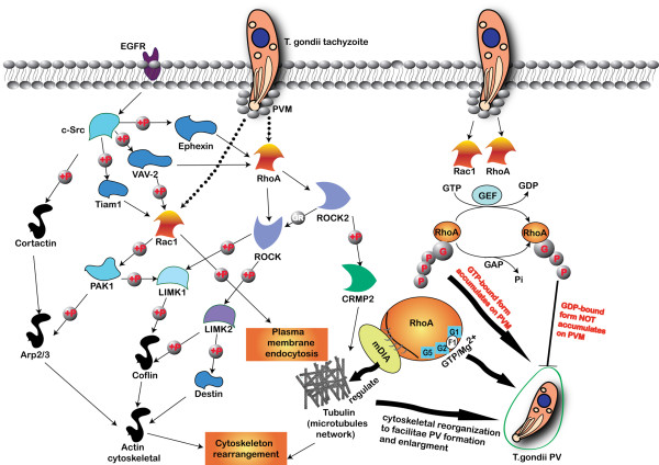
Figure 8.
Cell signaling related to RhoA and Rac1 regulated cytoskeleton reorganization in T. gondii infection. c-Src is activated by EGF induced EGF receptor activation and followed by Ephexin, VAV-2 and Tiam 1 phosphorylation. Ephexin phosphorylation promotes its GTPase activity toward RhoA and ROCK. ROCK directly phosphorylates LIMK1 and LIMK2, which in turn phosphorylate destrin and cofilin. ROCK2 phosphorylates CRMP2, and CRMP2 phosphorylation reduces its tubulin-heterodimer binding and the promotion of microtubule assembly. Activation of VAV-2 activates RhoA and Rac1. In the downstream of Rac1, p21-activated kinase 1 (PAK1) activates LIMK1 and regulates the actin cytoskeletal reorganization through the phosphorylation of the actin-depolymerizing factor cofilin and destrin. PAK1 also phosphorylates Arp2/3 complex to promote actin polymerization. Cortactin is a prominent target of c-Src, and regulates cytoskeletal dynamics. Tyrosine phosphorylation of cortactin reduces its F-actin cross-linking capability. In our research, we are not clear about the upstream of the RhoA and Rac1 GTPases cell signaling involved in T. gondii infection, but we can see the activation of RhoA and Rac1 of host cells and the reorganization of the cytoskeleton for PV formation. RhoA and Rac1 GTPases accumulate on the PMV regardless of the parasitic strain virulence, and the accumulation is dependent on their GTPase activity. The recruited RhoA or Rac1 on the PVM are probably in GTP-bound active form. The RhoA GTPase is recruited to the PVM as soon as the T. gondii tachyzoite invaded the host cell either through the host cell membrane or from the cytosol. The decisive domains for the RhoA accumulation on the PVM includes the GTP/Mg2+ binding site (F1), the mDia effector interaction site, the G1 box (G1), the G2 box (G2) and the G5 box (G5). The reorganization of host cell cytoskeleton facilitates the formation and enlargement of T. gondii PV in the host cell.

