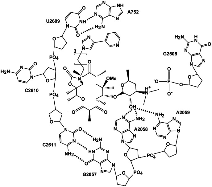
Figure 1. 2D representation of telithromycin and bases within the macrolide binding pocket showing the three biologically relevant telithromycin-ribosome interactions that are the subject of this study: 2′-OH to 2058/2059 hydrogen bonding, stacking between telithromycin's ARM and A752-U2609 WC base pair, and ionic interactions between telithromycin's 3′-protonated dimethylamine and G2505.
Dotted lines represent hydrogen bonding. Hydroxyl groups of the ribose sugars are omitted for clarity.

