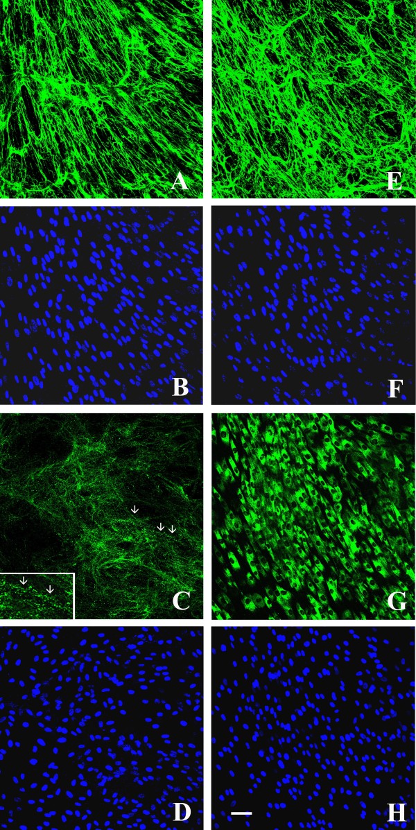
Figure 3.
Confocal laser microscopy of the collagen VI matrix in control and UCMD-confluent dermal fibroblasts after 4-days of L-ascorbic acid treatment.In vivo staining of the collagen VI network in control fibroblasts detected a normal quantity and a parallel alignment of collagen VI microfibrils (a), whereas UCMD cells secreted a decreased amount of collagen VI (c), and microfibrils showed a punctuate and discontinuous pattern of deposition (arrows, inset). In fixed and permeabilized samples, collagen VI immunofluorescence detected a significant intracellular retention in the majority of UCMD fibroblasts (g), whereas control cells exhibited a normal collagen VI deposition (e). Cell density was checked by counterstaining with Hoechst (b, f, d, h). Scale bar: 50 μm.

