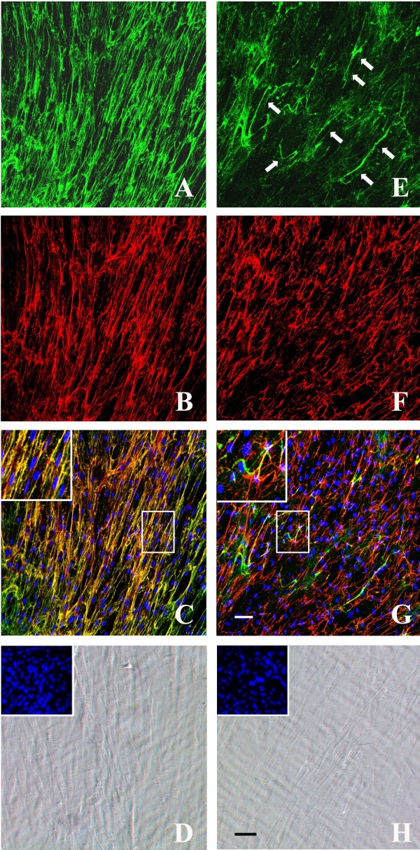
Figure 4.
Double immunolabelling with collagen VI and fibronectin antibodies. Double immunolabelling with collagen VI (green) and fibronectin (red) antibodies of the extracellular matrix secreted by cultured dermal fibroblasts from control (a-c) and UCMD patient (e-g) after 10 days of ascorbate treatment. Phase contrast images of control (d) and UCMD (h) fibroblasts showed the state of cell confluence (nuclei were stained with Hoechst, in insets). In control cells, collagen VI (a) and fibronectin (b) networks showed a similar distribution pattern and colocalized in several points, as revealed by their merge signal (yellow, high magnification in the inset). UCMD fibroblasts secreted a decreased amount of collagen VI matrix (e) with rare bundles of short microfilaments (e, arrows). UCMD cells produced an altered fibronectin network with short and irregularly aligned fibrils (f), and the colocalization with the collagen VI matrix was significantly impaired (g, high magnification in the inset). Nuclei were stained with DRAQ5 (pseudocolored in blue in c and g). Scale bar: 50 μm.

