
Figure 2. Ultrasonography findings showing a normal and thickened gallbladder wall in dengue patients.
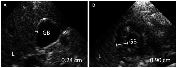
Handheld ultrasonography performed at the bedside with the patient in supine position during examination. (A) normal gallbladder wall (<0.30 cm) (B) and thickened gallbladder wall. The gall bladder wall is thickened with 0.90 cm and has a subserosal fluid layer, giving the gallbladder wall a multiple layer aspect. Abbreviations: L: liver; GB: gallbladder lumen.