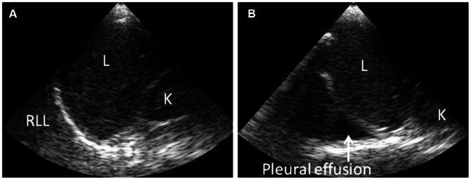
Figure 3. Ultrasonography findings showing presence or absence of pleural effusion in dengue patients.

Handheld ultrasonography at the bedside with patient in supine position during examination. (A) Normal right costophrenic angle. (B) Pleural effusion in the right costophrenic angle (arrow). Abbreviations: L: liver; K: kidney, RLL: right lower lobe of lung.
