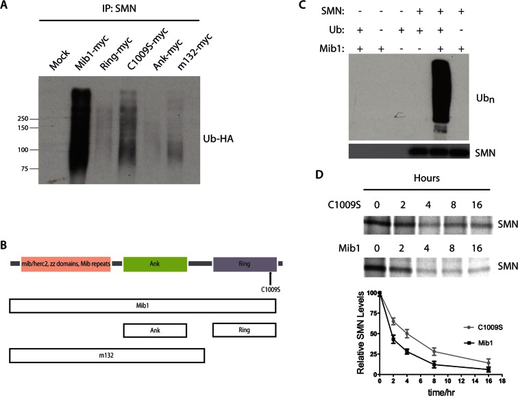
FIGURE 1:
(A) NSC-34 cells were transfected with 2 μg Mib1 and 1 μg HA-Ub cDNAs. The cells were harvested 48 h later, and endogenous SMN was immunoprecipitated. Immunoprecipitated proteins were resolved by SDS–PAGE, and the proteins were analyzed by Western blotting. The blots were probed with an HA antibody to detect ubiquitinated SMN. (B) Schematic representation of Mib1 protein domains. (C) Cell-free SMN ubiquitination assay. Recombinant SMN was incubated with E1 and E2 (UBCH5B) enzymes with or without Mib1 and ubiquitin for 1 h at 37°C. Western blots were probed with an anti-polyubiquitin antibody (FK1). (D) Pulse–chase analysis of endogenous SMN in the presence of 2 μg Mib1-myc or Mib1-C1009S-myc. The data represent mean ± SEM of three independent experiments.

