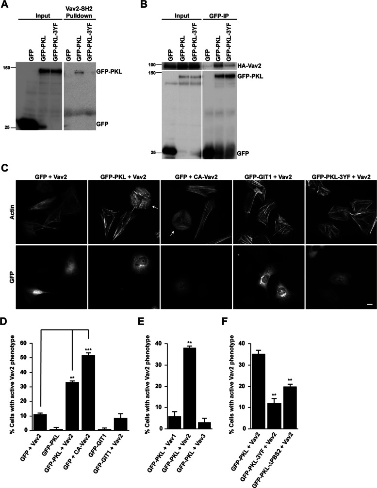
FIGURE 1:
PKL and Vav2 coexpression induces an “active” Vav phenotype. (A) Representative Western blot showing GFP-PKL binding to GST-Vav2-SH2 domain. In contrast, the nonphosphorylatable GFP-PKL-3YF does not bind. (B) Representative Western blot showing coimmunoprecipitation of HA-Vav2 with GFP-PKL. (C) Fluorescence analysis of cells expressing GFP, GFP-PKL, or GFP-GIT1 together with HA-Vav2 or HA-CA-Vav2 as indicated. Arrows indicate transfected cells exhibiting “active” Vav2 phenotype. Scale bar, 10 μm. (D–F) Cell counts of cells coexpressing the constructs indicated. GFP-positive cells exhibiting an “active” Vav2 phenotype were scored. At least 100 cells per condition were counted, and numbers are mean values ± SEM for three separate experiments. Significance was determined using a Student's t test. **p < 0.005, ***p < 0.0005.

