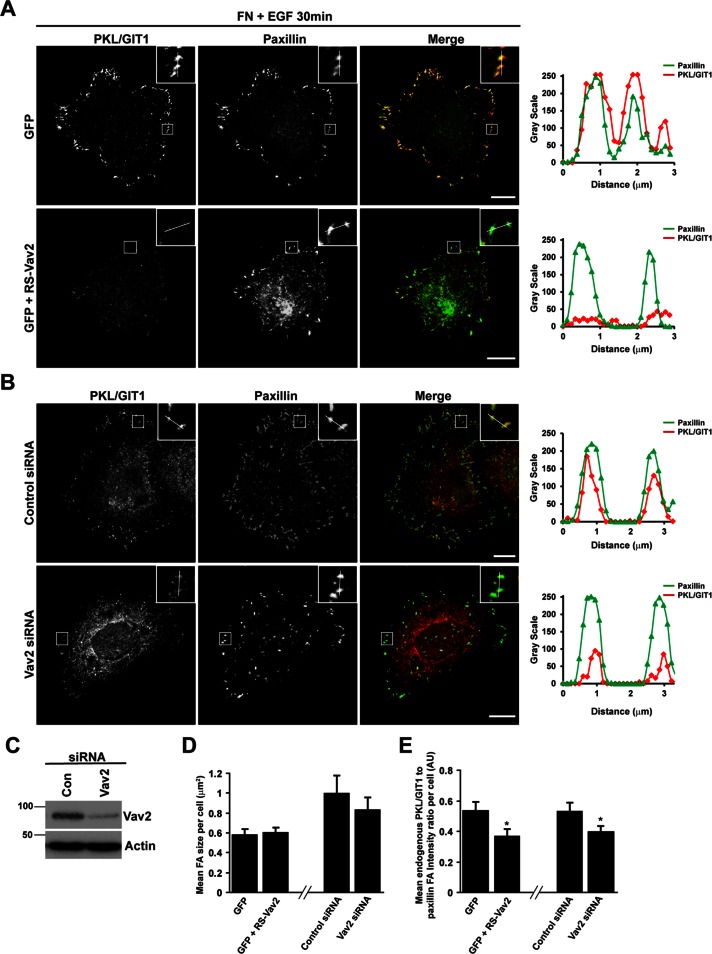
FIGURE 6:
Vav2 activity is required for EGF-stimulated localization of PKL to adhesions. (A) HT1080 cells transfected with GFP or GFP plus dominant-negative L342R/L343S Vav2 (RS-Vav2), which lacks nucleotide exchange activity, were spread on FN for 30 min in the presence of EGF and then stained for PKL/GIT1 and paxillin. Images are contrast enhanced to equal degrees for presentation. Scale bars, 10 μm. (B) HT1080 cells were transfected with either control siRNA or siRNA targeting Vav2. After 72 h cells were spread on FN for 30 min in the presence of EGF and then stained for PKL/GIT1 and paxillin. Images are contrast enhanced to equal degrees for presentation. Scale bars, 10 μm. Line profiles through adhesions indicated in A and B demonstrate decreased intensity of PKL in paxillin-positive adhesions. (C) Representative blot showing efficient knockdown of Vav2 in HT1080 cells. The average focal adhesion size (D) and the average ratio of PKL/GIT1 intensity to paxillin intensity in adhesions per cell (E) were quantified in background-subtracted raw images using ImageJ. Values are means ± SEM for three experiments and at least 10 cells per experiment. Significance was determined using Student's t test. *P < 0.05.

