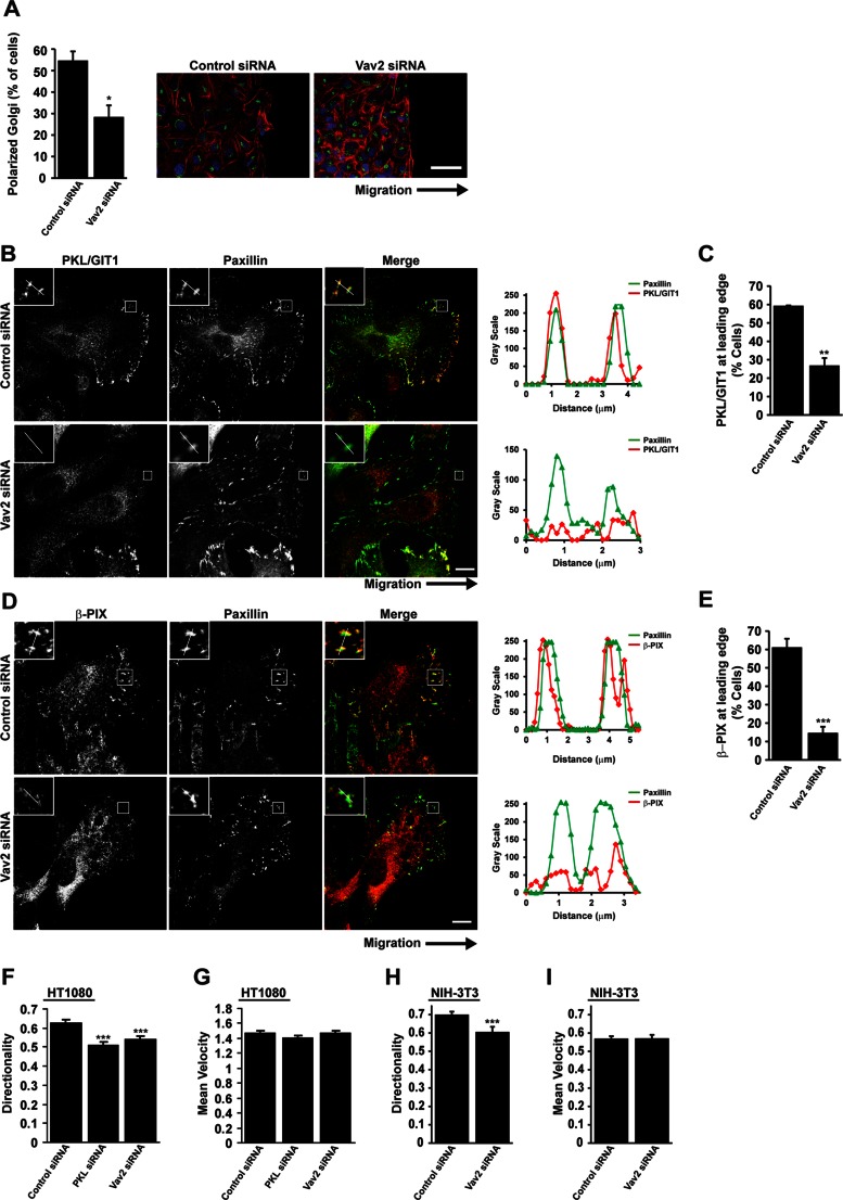
FIGURE 9:
Vav2 is required for localization of PKL/GIT1 and β-PIX to the leading edge of migrating cells. (A) HT1080 cells were transfected with siRNAs targeting Vav2. After 72 h cells were replated to form a confluent monolayer and wounded. Cells were allowed to migrate into a scrape wound for 2 h before fixation and staining for the Golgi marker GM130, paxillin, DAPI, and rhodamine–phalloidin. Quantitation of polarized Golgi at the wound edge demonstrates a significant decrease after Vav2 knockdown. *p < 0.05. Scale bar, 50 μm. (B, D) HT1080 cells depleted of Vav2 show perturbed localization of PKL/GIT1 and β-PIX to the leading edge of migrating cells. Scale bars, 10 μm. Line profiles through adhesions indicated in B and D demonstrate decreased intensity of PKL/GIT1 and β-PIX in paxillin-positive adhesions at the leading edge. (C, E) Quantitation of the number of cells at the wound edge, demonstrating polarized distribution of PKL/GIT1 and β-PIX. Values are means ± SEM for three experiments and at least 30 cells per experiment. Significance was determined using Student's t test. **p < 0.05, ***p < 0.005. (F, G) HT1080 or (H, I) NIH 3T3 cells were transfected with siRNA targeting PKL or Vav2 as indicated. After 72 h cells were replated to form a confluent monolayer and wounded. Cell migration was allowed to proceed for 16 h and individual cells subsequently tracked using ImageJ. Values for directionality (F, H) and velocity (G, I) were determined for at least 40 cells per experiment and three separate experiments. ***p < 0.0005.

