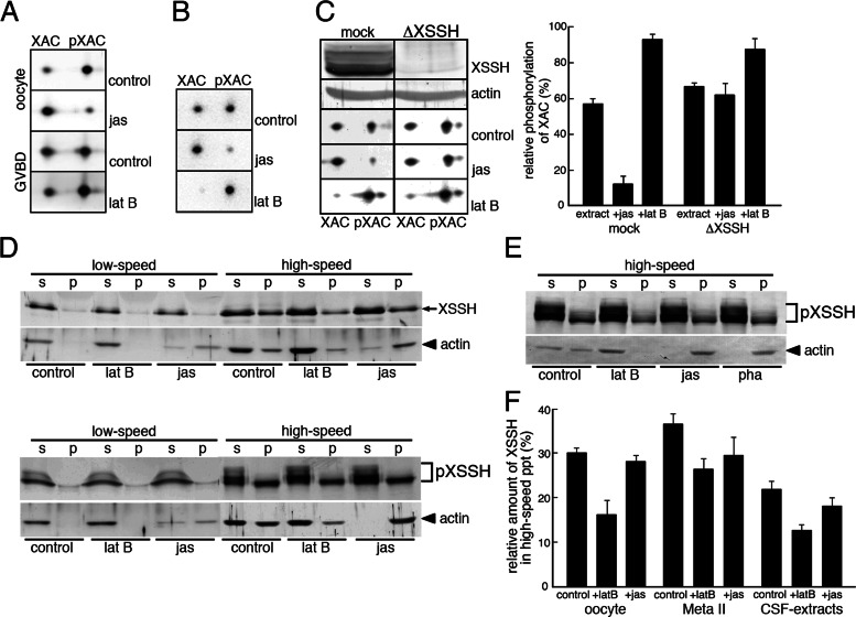
FIGURE 4:
Effects of F-actin levels on the XAC phosphatase activity of XSSH. (A) Phosphorylation states of XAC were examined by 2D immunoblots of XAC in immature oocytes (oocyte) and maturing oocytes just after GVBD, which were treated with jasplakinolide (jas) or latrunculin B (lat B). Control indicates the treatment with vehicle alone. (B) Phosphorylation states of XAC were examined by 2D immunoblots of XAC in CSF extracts in the presence of jas or lat B. Control indicates addition of vehicle alone. (C) Phosphorylation states of XAC were examined by 2D immunoblots of XAC in mock-depleted CSF extracts (mock) or XSSH-depleted CSF extracts (ΔXSSH) in the absence (control) or presence of jas or lat B. XSSH was sufficiently depleted from CSF extracts (front row, XSSH) as judged by the anti-actin blots (second row, actin). The spots of XAC and pXAC were quantified by densitometry, and percentages of pXAC are shown. (D) Immature oocytes (top) and maturing oocytes just after GVBD (bottom) treated with vehicle alone (control), lat B, or jas were centrifuged at 12,000 × g for 15 min (low speed), and the supernatants were further ultracentrifuged at 436,000 × g for 20 min (high speed) to separate the supernatants and pellets. The supernatants (s) and pellets (p) were subjected to SDS–PAGE and examined by immunoblotting with anti-XSSH or anti-actin antibody. (E) Immunoblots of XSSH and actin in high-speed supernatants (s) and pellets (p) of CSF extracts treated with vehicle alone (control), 25 μM lat B, 10 μM jas, or 10 μM phalloidin (pha) as described in D. (F) The XSSH bands in high-speed supernatants and precipitates (ppt) were quantified by densitometry, and percentages of precipitated XSSH are shown. Bars, mean + SE from three independent experiments using immature oocytes (oocyte), mature oocytes (Meta II), and CSF extracts in the absence (control) or presence of latrunculin B (+latB) or jasplakinolide (+jas).

