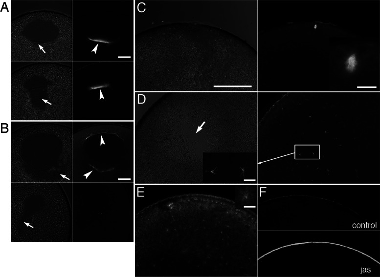
FIGURE 6:
Effects of injection of anti-XSSH antibody on assembly of MTOC-TMA and meiotic spindles. (A, B) Phase contrast (left) and immunofluorescence (right) microscopy of control oocytes injected with buffer alone (A) or oocytes injected with anti-XSSH antibody (B). Microtubules were stained with anti-tubulin antibody (right). Assembly of MTOC-TMA (arrowhead in A, top right) at the basal region of GV and its migration toward the animal pole (arrowhead in A, bottom right) are clearly visible in control (A), whereas its structure is faint in antibody-injected oocytes (arrowheads in B, top right). The yolk-free region formed at the basal region of GV (indicated by arrows in phase contrast micrographs) showed broader and irregular shape in anti-XSSH antibody-injected oocytes (B). (C, D) Phase contrast images (left) and immunofluorescence staining for tubulin (right) of control oocytes 4 h after GVBD (C) and antibody-injected oocytes fixed at 4 h after control oocytes formed WMS (D). Meiotic spindles were already assembled in control oocytes (C, magnified in inset), whereas fine and radial microtubule structures were formed (D, left, magnified) and scattered around the yolk-free region in antibody-injected oocytes (D, arrow). (E) A jasplakinolide-treated oocyte was fixed at 4 h after control oocytes formed WMS and stained with anti-tubulin antibody. An example of radial microtubule structures is shown in the inset on the upper right corner. (F) Staining of control (top) and jasplakinolide-treated (bottom) oocytes for actin filaments by anti-actin antibody. Note the cortical accumulation of actin filaments by jasplakinolide treatment. Bars, (A, B) 100 μm, (C, applies to C– F) 200 μm, (insets) 20 μm.

