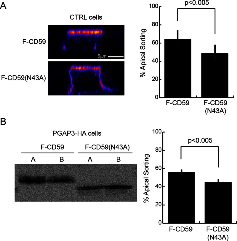
FIGURE 5:
N-glycosylation is required for proper sorting of fully remodeled GPI-APs and lysoGPI-APs. (A) Orthogonal views of control cells expressing F-CD59 or F-CD59(N43A) grown on filters for 6 d. The signal intensity was measured by immunofluorescence staining of F-CD59 after fixation in 100% methanol at −20°C for 20 min and labeling with an anti-CD59 antibody. The means of apical sorting (percentage of apical signal/total signal) + SD for 20 cells from two independent experiments are shown. The micrographs are pseudocolored as in Figure 4B. (B) Polarized release of F-CD59 or F-CD59(N43A) into the apical (A) and basolateral (BL) chambers of PGAP3-HA cells grown on filters for 6 d during a 2-h chase after pulse labeling was analyzed using anti-FLAG M2 beads. The means of apical sorting (percentage of apical signal/total signal) + SD from five independent experiments are shown.

