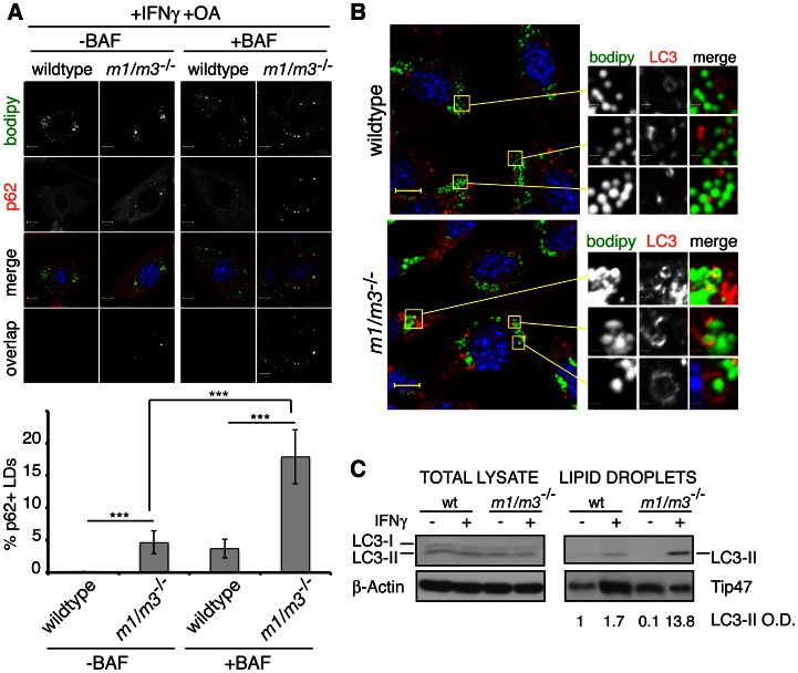
Figure 8. IRGM-deficient LDs recruit p62 and LC3-II.
(A and B) Wildtype and Irgm1/m3−/− MEFs were treated overnight with OA and IFNγ and stained with BODIPY and anti-p62 or anti-LC3, respectively. Representative images of at least 3 independent experiments are shown. Where indicated, cells were treated with Bafilomycin (BAF). (A, lower panel) Quantitative analyses of p62 co-localization with LDs were done using MBF-ImageJ software as described in Materials and Methods (***, p<0.005). (C) Total cell lysates and LD protein preparations from the indicated samples were analyzed for LC3 protein expression. Densitometric analyses for protein quantification were carried out using ImageJ 1.45 s software. The relative optical density (O.D.) for LC3-II expression normalized against expression of the LD marker Tip47 is listed below the corresponding lanes in arbitrary units.

