
Figure 4. Treg accumulation and disease progression in TLR2KO mice is not altered by the transfer of WT Tregs.
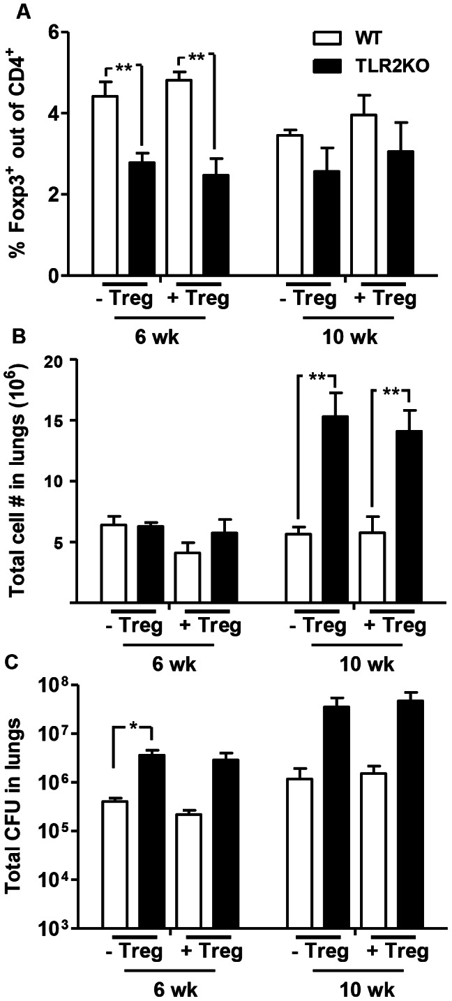
CD4+CD25+ Tregs purified from naïve WT mice were transferred to TLR2KO and WT control mice one day prior to and 4 weeks after Mtb infection (0.5×10⁶ cells per mouse for each transfer). Non-recipient WT and TLR2KO control mice were infected at the same time. Single cell suspensions were prepared at the indicated time points after Mtb challenge. Lung cells were stained with antibodies against CD4 and Foxp3, followed by flow cytometric analysis. Expression of Foxp3 is presented as a percentage of the CD4+ cell population (A). Total number of viable cells in the lungs was determined by trypan blue exclusion method (B). Bacterial burden in the lungs was determined by plating serial dilutions of lung homogenates onto 7H11 agar plates (C). Data include 4–5 mice per group and are presented as mean ± SEM. *, p<0.05; **, p<0.01.