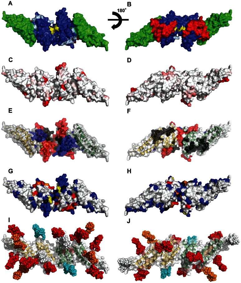
Figure 1. Design of surface re-engineered PvDBPII recombinant proteins containing additional N-linked glycan residues.
(A–B) Subdomains and DARC binding models. Subdomain 1(red), subdomain 2 (blue), and subdomain 3 (green). Critical binding residues for model 1 are colored light blue and for model 2 are colored yellow. (C–D) Fractional Shannon entropy values [70] from 0.000 (white) to 0.243 (red) for sequence polymorphism over the PvDBPII surface as compared with maximally entropic distribution over all amino acids. (E–F) Epitopes recognized by blocking antibodies [20]; black (low inhibitory), blue (medium inhibitory), red (high inhibitory). (G–H) Meta-analysis of mutations that reduce or do not affect the PvDBP-DARC interaction: blue residues (no effect); yellow residues (minor); orange residues (moderate); red residues (major); black residues, differences between studies (see Table S1). (I–J) Location of engineered N-glycosylation sites modeled as high mannose forms; white (wild type), cyan (STBP glycan), Orange (P1 and Max), red (Max). All images modeled in PyMol on the PvDBPII dimer structure [13], viewing opposite ends of the dimeric two-fold axis for subpanels I and II; missing density in the crystal structure has been left missing, though it contains polymorphic, inhibitory, antibody recognition and glycan-bearing sites. PvDBPII monomers are colored yellow and green in panels E, F, I, and J.

