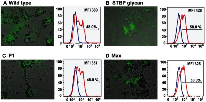
Figure 4. Functional characterization of hyperglycosylated PvDBPII variants by COS-7 cell-RBC binding assay.
PvDBPII wild type (A), STBP glycan mutant (B), P1 (C) and Max (D) DBPII glycosylation variants were expressed as a GFP fusion protein in COS-7 cells and incubated with 0.5% hematocrit erythrocytes. GFP-positive transfected cells with five or more RBC (rosettes) were considered positive for binding. The histogram shows the percentage surface labeling when GFP-positive transfected cells were probed with pre-immune (blue) or anti-PvDBPII plasma (red). The gating strategy for GFP-positive and anti-PvDBPII positive is shown in Figure S3. The percentage signifies GFP-positive cells that are labeled with immune plasma, indicating surface expression of PvDBPII and DBPII glycosylation variants. The shift in mean fluorescence intensities between pre-immune and immune plasma is indicated as MFI.

