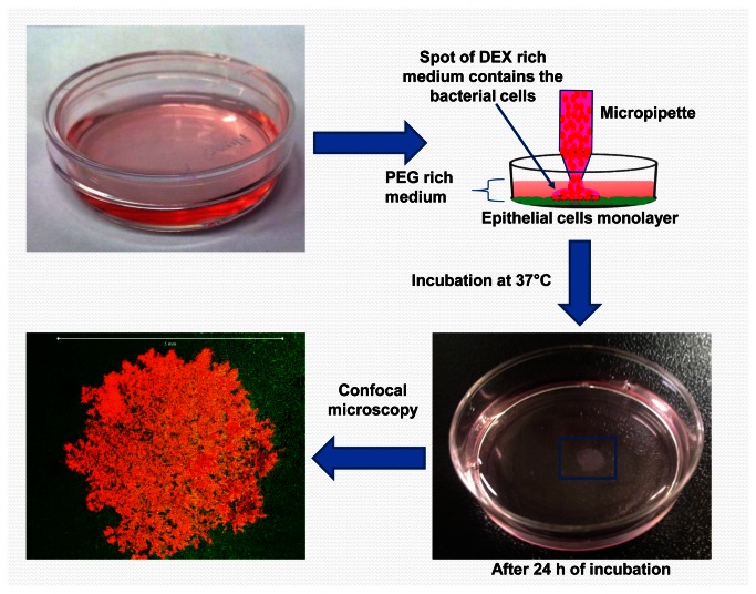
Figure 1. A schematic diagram shows the procedure of making ATPS derived bacterial communities on epithelial monolayer using the first ATPS formulation.
The mammalian cells (MCF10a cell line) were grown in supplemented DMEM/F12 medium inside a 35 mm Petri dish as usual until it formed a monolayer sheet (~80% confluency). The growth medium was then removed and replaced with 2.5 ml of PEG rich medium. This is followed by spotting drops of the DEX rich medium containing the bacteria (E. coli/pHKT3) using a conventional micropipette. After 24 h of incubation at 37°C, the resulting bacterial community is visible and can be analyzed microscopically (scale bar: 5 mm).

