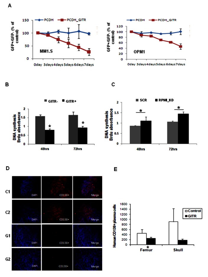
Figure 3. Effect of GITR on MM tumor proliferation in vitro and in vivo.
A) MM cells (MM.1S and OPM1) were transfected with PCDH empty and PCDH-GITR with GFP labeled lentiviral vector (Cat# CD511B-1, SBI Inc.) respectively. Cell proliferation has been evaluated by using GFP competition assay. Expression of GFP and GITR has been examined by flow cytometry by using anti-human GITR PE labeled primary antobidy. GFP+ and GFP-MM cell were sorted by BD laser II flow machine. Ratio of GFP+/GFP-was recorded everyday after combination. B) Effect of GITR-overexpression on MM1.S cell line proliferation. 96 wells plate coated with MM1.S cells were read after 48 and 72 hours respectively. Anti-Brdu-pod was used to detect the absorbance. Mean±SD. *, P<0.05. C) Effect GITR-knockdown on RPMI.8226 cell line proliferation (RPMI.8226). 96 wells plate coated with MM1.S cells were read after 48 and 72 hours respectively. Anti-Brdu-pod was used to detect the absorbance. Mean±SD. *, P<0.05. D) SCID mice were injected i.v. with 5 million MM1.S cells, transfected with either empty vector (contrl C1, C2) or GITR (GITR+, G1, G2). In vivo tumor progression has been evaluated by using immunofluorescence staining with anti-human CD138 monoclonal antibody after 4 weeks injection, on bone marrow femurs. E) Detection of MM cells from tissues of mice injected with either empty vector (control) or GITR (GITR+). MM cells have been detected by using flow cytometry analysis for CD138. Mean±SD. *, P<0.05; **, P<0.01.

