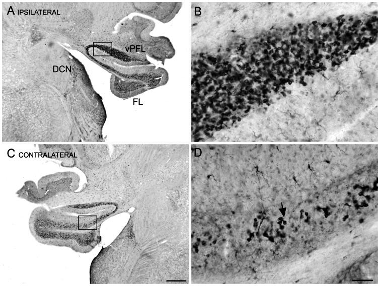
Figure 1. Doublecortin (DCX) immunoreactivity (IR) in UBCs of the DCN and cerebellum ipsilateral to the exposure.
A. Photomicrograph through the brainstem and cerebellum showing DCX-IR neurons. The rectangle shows the location of the higher magnification photomicrograph in B. B. DCX-IR in UBCs of the vPFL. C. DCX-IR neurons in the vPFL, FL and DCN on the side contralateral to the exposure. The rectangle shows the location of the photomicrograph in D. Scale bar 500 µm. D. DCX-IR neurons are UBCs, example at arrow. Scale bar 50 µm. Abbreviations: DCN, dorsal cochlear nucleus; FL, flocculus; UBC, unipolar brush cell vPFL. ventral paraflocculus.

