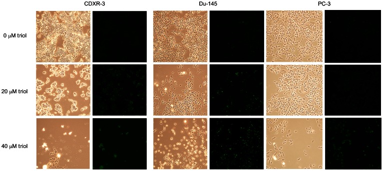
Figure 5. Triol induces apoptosis in prostate cancer cells.
LNCaP CDXR-3, DU-145, and PC-3 cells were treated with 0, 20, and 40 µM triol for 48 hrs. Cell morphology was determined by light microscopy. TUNEL assay was performed as described in Materials and Methods to determine the apoptotic cell population. Green fluorescent light indicated apoptotic cells stained with TUNEL assay. Images were viewed at 200× with Olympus confocal microscope.

