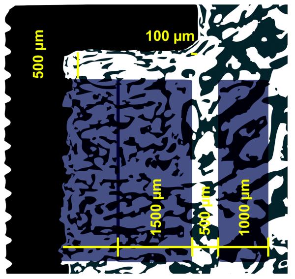
Fig. 3.
Histomorphometry – The region of interest (ROI) is defined on both sides of the implant. The ROIs are illustrated on one side of the implant above. Tissue ongrowth (surface fraction) at the surface of the implant, the tissue volume (volume fraction) in the grafted gap of 2.5 mm (divided into an outer region (1500 μm) and inner region reaching the surface of the implant) and tissue volume in region of intact non-implanted bone (1000 μm) are defined. The regions were defined from the end-washer margin as a fixed point with a 100 μm clearance at the gap-intact-bone-interface and 500 μm below the washer. ROI were defined at magnification × 1.25. Total assessment magnification × 30.3. For histomorphometric assessment, the corresponding magnifications were × 20 and × 402.

