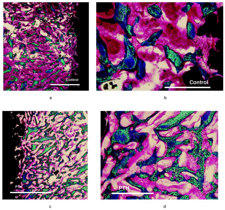
Fig. 5.
Photomicrographs of representative histological samples showing a-b) control and c-d) parathyroid hormone (PTH). Left images a-c) bar 2000 μm and right images b-d) bar 500 μm. Staining technique 0.4% basic fuchsin (red) and 2% light green (green = bone) and black = implant. PTH shows increase in bone in the morsellised impacted gap with numerous connective trabeculae of wo ven bone and elements of bone allograft. In the control group bone graft appears with sparse new bone.

