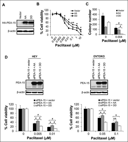
Figure 2.
Overexpression of PEA-15 phosphorylated at both Ser104 and Ser116 enhanced the sensitivity of ovarian cancer cells to paclitaxel. A, Expression of HA (hemagglutinin)-tagged PEA-15 was determined by western blotting in SKOV3.ip1 stable cells. β-actin served as a loading control. The sensitivity of SKOV3.ip1 stable cells to paclitaxel was determined by (B) WST-1 assay 72 hours after paclitaxel treatment and (C) anchorage-independent growth assay 3 weeks after paclitaxel treatment. In A-C, vector, SKOV3.ip1-vector; AA, SKOV3.ip1-AA (stably expressing nonphosphorylatable PEA-15 at Ser104 and Ser116, which were substituted with alanine); DD, SKOV3.ip1-DD (stably expressing phosphomimetic PEA-15 at Ser104 and Ser116, which were substituted with aspartic acid). D, PEA-15 expression in HEY and OVTOKO cells was silenced using PEA-15-targeting siRNA, and then cells were transiently transfected with the pcDNA3.1 vector, the pcDNA3.1 vector carrying nonphosphorylatable PEA-15 (AA; Ser104 and Ser116 of PEA-15 substituted with alanine), or the pcDNA3.1 vector carrying phosphomimetic PEA-15 (DD; Ser104 and Ser116 of PEA-15 substituted with aspartic acid). Expression of PEA-15 was determined by western blotting 48 hours after transfection (upper panels). β-actin served as a loading control. Following transfection, the sensitivity of HEY and OVTOKO cells to paclitaxel was determined by trypan blue exclusion assay 72 hours after paclitaxel treatment (lower panels). *, P < 0.05; **, P < 0.01; #, P < 0.001.

