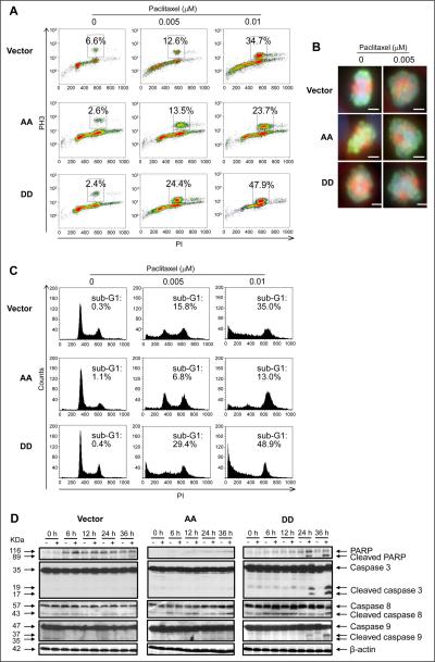
Figure 3.
Overexpression of PEA-15 phosphorylated at both Ser104 and Ser116 promoted induction of mitotic arrest and apoptosis by paclitaxel. SKOV3.ip1 stable cells were treated with paclitaxel and then subjected to immunofluorescence staining with anti-phosphohistone H3 antibody and analyses by (A) flow cytometry 12 hours after paclitaxel treatment and (B) fluorescence microscopy 6 hours after paclitaxel treatment. In A, the x-axis indicates the intensity of propidium iodide (PI) staining, and the y-axis indicates the intensity of phosphohistone H3 (PH3) staining. In B, scale bar, 100 μm. C, SKOV3.ip1 stable cells were treated with paclitaxel for 36 hours and then subjected to cell cycle analysis using PI staining and flow cytometry. The x-axis indicates the intensity of PI staining, and the y-axis (counts) shows the cell number. D, SKOV3.ip1 stable cells were treated with paclitaxel for 36 hours and then subjected to western blotting to verify cleavage of PARP and caspases 3, 8, and 9. β-actin served as a loading control. Vector, SKOV3.ip1-vector; AA, SKOV3.ip1-AA (stably expressing nonphosphorylatable PEA-15 at Ser104 and Ser116, which were substituted with alanine); DD, SKOV3.ip1-DD (stably expressing phosphomimetic PEA-15 at Ser104 and Ser116, which were substituted with aspartic acid).

