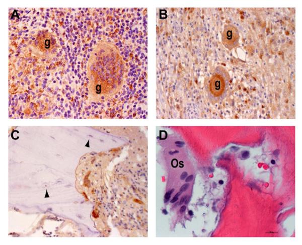
Figure 5. Evidence of macrophage differentiation and bone-remodeling.

CD68 immunostaining from an HIV-negative (A) and two HIV-positive (B, C) specimens. CD68+ individual macrophages (arrows) and multinucleated giant cells (g), as well as CD68− osteocytes (arrowheads) are indicated (×40 magnification). D) H&E staining depicts a multinucleated osteoclast (Os) in close proximity to damaged bone from an HIV-negative patient (×80 magnification).
