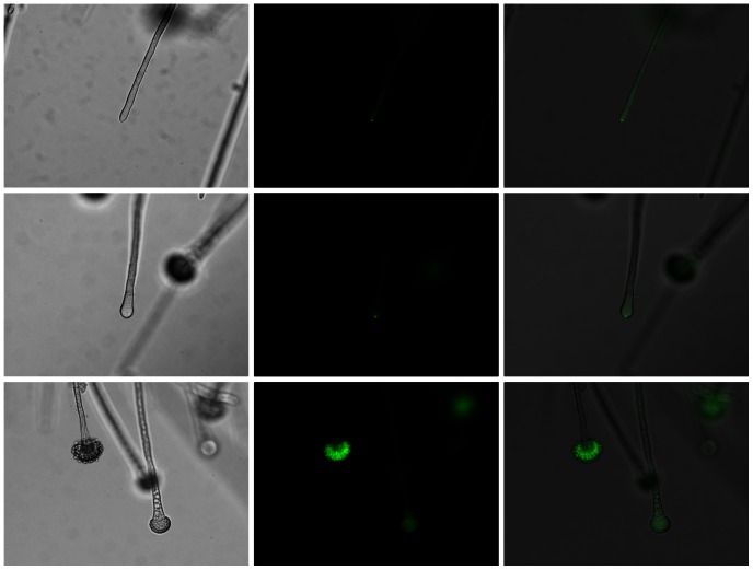
Figure 5. GFP-SrgA localizes to conidiophores.
GFP-SrgA localizes to the apex of both hyphae and conidiophores. A punctate accumulation at the tip is seen in both hyphae and the early stages of vesicle swelling (top and middle rows, respectively), but a more diffuse localization is evident in mature conidiophores (bottom row). Left column: brightfield; middle column: GFP fluorescence; bottom column: Merged image.

