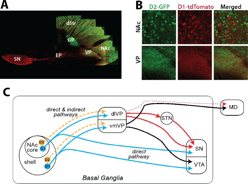
Figure 2.
Output pathways of NAc D1-MSNs and D2-MSNs. A. Micrograph of projections from D1-(red) and D2-expressing cells (green) in D1-tdTomato x D2-GFP transgenic mice demonstrates that both MSN types in nucleus accumbens (NAc) project to ventral pallidum (VP), while D1-MSNs also terminate in midbrain (substantia nigra, SN, shown in this section, and ventral tegmental area, VTA, not shown). In contrast, dorsal striatal (dStr) D1-MSNs and D2-MSNs send distinct projections through the direct and indirect pathways. Output to the entopeduncular nucleus (EP) and SN is entirely from D1-MSNs, whereas the indirect projection to the globus pallidus (GP) is from D2-MSNs. B. While minimal co-localization is observed in NAc D1-MSNs and D2-MSNs, both MSNs terminate directly in VP. C. Diagram illustrating the overlap of D1- and D2-MSN projections to VP, with the NAc core projecting to dorsolateral (dlVP) and the shell innervating the ventromedial compartment (vmVP). The dlVP projects to the subthalamic nucleus (STN) and SN (solid red lines), with minor contributions to mediodorsal thalamus (MD, dashed red line). The vmVP projects within the basal ganglia to VTA and out of the basal ganglia to MD (solid black lines). Unlike the NAc projection to VP, NAc innervation of the ventral mesencephalon is entirely a direct pathway and contains axons from D1-MSNs only.

