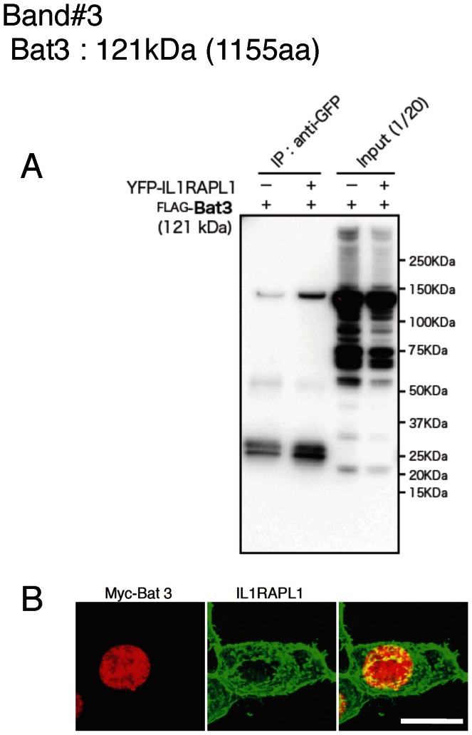
Figure 2. Analysis of IL1RAPL1-binding protein candidates (band #3).
A, Coimmunoprecipitation of YFP-IL1RAPL1 with FLAG-Bat3 in HEK 293T cells. Immunoprecipitation with anti-GFP antibody and total cell lysates, followed by western blotting with anti-FLAG antibody are shown. B, Colocalization of IL1RAPL1 (middle, green) and myc-Bat3 (left, red) in HEK 293T cells. Merged images are shown (right). Scale bar, 10 μm.

