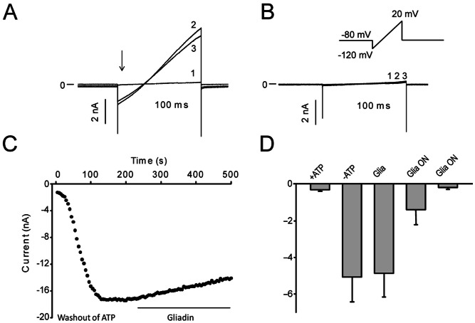
Figure 4. Gliadin digest incubation inhibits current through KATP.
Kir6.2 and SUR1 were expressed in HEK-293 cells. The cells were incubated overnight in either 300 µg/ml gliadin digest or a corresponding volume of enzyme mixture. Currents were activated by a ramp protocol every 5 s. A. Representative KATP currents in a cell incubated with the enzyme mixture overnight (1) prior to ATP washout, (2) after ATP washout in the presence of enzyme mix and (3) after 2.5 minutes exposure to gliadin digest. B. Representative currents (traces overlaid in plot) from a cell incubated with gliadin digest overnight, (1) prior to ATP washout, (2) after ATP washout in the presence of the enzyme mixture and (3) after application of the enzyme mixture for 2.5 minutes. C. The time-dependence of the effect of gliadin digest on KATP currents. Representative data from a cell incubated in enzyme mix overnight. The data points represent maximal inward current during washout of endogenous ATP and after application of gliadin digest. D. Summarized current densities in : +ATP, average current density prior to ATP washout in cells incubated in enzyme mix overnight (n = 7), −ATP, average current density after washout (n = 9). Gliadin digest: average current density after 2.5 min exposure to 300 µg/ml gliadin digest (n = 9). The effect of gliadin digest was not significant. In 10/12 cells incubated in gliadin digest overnight there was no expressed KATP current. In the first bar graph all cells are included; in the second bar graph the 2 cells with current are excluded. For cells incubated in the enzyme mixture, 3/12 cells had no current and were excluded from the analysis.

