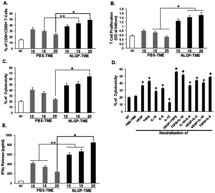
Figure 5. NLGP normalizes T cell functions within TME.
Lysates were prepared from tumors of PBS and NLGP treated mice, designated as PBS-TME and NLGP-TME. MNCs from normal mice were incubated with PBS-TME and NLGP-TME, prepared from tumors of different days for 120 hrs. A. percentage of CD8+CD69+ T cells was analyzed within different TME treated MNCs * p<0.01, ** p<0.05. B. CD8+ T cells were then allowed to proliferate for 72 hrs and proliferation was determined by MTT assay. NLGP stimulation in MNCs was kept as control. *p<0.01. C. After 120 hrs of incubation with different TMEs, CD8+ T cells were purified by MACS. Cytotoxicity of these cells towards sarcoma 180 cells was assessed by LDH release assay. ▴ p<0.01. D. Different growth factors (VEGF, TGFβ) and cytokines (IL-10, IL-6 and IL-12) in single or in combination were neutralized within PBS-TME (Tumor lysate prepared from tumor of day 20) using their respective antibodies. Splenic MNCs were then exposed to differentially neutralized PBS-TME and after 120 hrs incubation CD8+ T cells were purified by MACS. Cytotoxicity of these differentially exposed CD8+ T cells towards Sarcoma 180 cells was measured. • p<0.001, + p<0.01 in comparison to PBS-TME. E. CD8+ T cells purified in similar fashion as described in C and cultured for 48 hrs. Cell supernatants were used to measure IFNγ level by ELISA. NLGP stimulation in normal CD8+ T cells was kept as control. p = 0.0023. In every case, comparison was made between PBS-TME exposed T cells vs same exposed to NLGP-TME on day 15 and 20, ▴p<0.01, **p<0.05.

