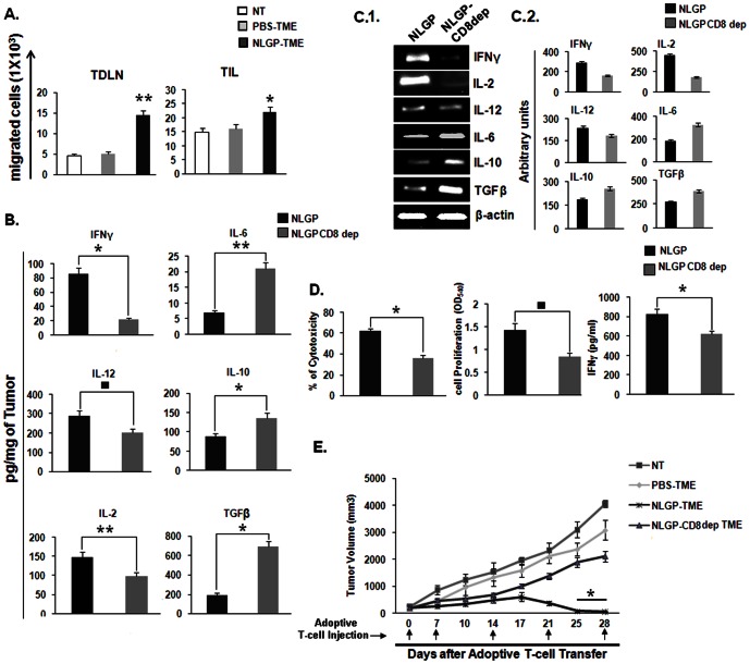
Figure 7. NLGP enhances T cell migration to TDLN and TIL to effectively kill tumors in vivo.
A. MNCs were isolated from normal mice and exposed to PBS-TME and NLGP-TME for 120 hrs, along with a set of unexposed cells. Then CD8+ T cells were purified by MACS. Purified cells were labeled with CFSE and injected into three groups of mice having tumors of identical volume. After 24 hrs migration of CD8+CFSE+ cells were detected in TDLN and TIL, **p<0.001, *p = 0.024. B. Depletion of CD8 impairs NLGP mediated TME normalization. Tumor tissue lysates, representing TME from either NLGP or CD8 depleted NLGP treated mice (n = 4 in each case) were assessed for IFNγ, IL-2, IL-12, IL-6, TGFβ and IL-10 by ELISA. Cytokines were quantitated as pg/mg of tumor tissue ± SE , *p = 0.009, **p = 0.008, ▪ p = 0.005, in comparison to PBS treated tumor on day 20. C.1. Total RNA was isolated from tumor of PBS and NLGP treated mice (n = 4 in each case) to analyze genes of IFNγ, IL-2, IL-12, IL-6, TGFβ and IL-10 by RT-PCR. C.2. Densitometric analysis was performed in each case. D. MNCs were isolated from normal mice and exposed to NLGP-TME and CD8 depleted NLGP-TME for 120 hrs. Then CD8+ T cells were purified by MACS and T-Cell proliferation, IFNγ release and cytotoxicity towards Sarcoma180 were measured *p = 0.01, ▪ p = 0.007, E. MNCs were isolated from normal mice and exposed to PBS-TME, NLGP-TME and CD8 depleted NLGP-TME for 120 hrs, along with a set of unexposed cells. Then CD8+ T cells were purified by MACS and activated CD8+ T cells (1×10⁷ cells) were adoptively transferred through tail vein into four groups of tumor bearing mice (n = 6 in each group) once weekly as described in Materials and Methods (n = 3). Mean tumor volume ± SD and survivability are presented, *p<0.001.

